Long Bone Diagram Endosteum ~ General features of a LONG BONE - Biology 225 with Watson at McNeese State University - StudyBlue. 1 endosteum has cells known as endosteal. This endosteal surface is usually resorbed during long periods of malnutrition, resulting in less cortical thickness. Long bones are longer than they are wide and are the major bones of the limbs. Below is a 3d map of the skeletal system. Periosteum spongy bone compact bone red bone marrow yellow bone marrow epiphysis marrow cavity articular cartilage diaphysis diaphysis site of endosteum epiphyseal line epiphyseal line spongy bone epiphysis articular cartilage
Inside the diaphysis is the medullary cavity, which is filled with yellow bone marrow in an adult. A long bone has two parts: (b) in this micrograph of the osteon, you can clearly see the concentric lamellae and central canals. The diaphysis and the epiphysis (figure 6.3.1). Below is a 3d map of the skeletal system.
long bone diagram | timothyakeller | Flickr from c1.staticflickr.com The endosteum (plural endostea) is a thin vascular membrane of connective tissue that lines the inner surface of the bony tissue that forms the medullary cavity of long bones. Microscopic structure of long bone / 9 fishbone diagram templates to get started. The hollow region in the diaphysis is called the medullary cavity, which is filled. An epiphyseal disk of cartilage at the junction of the diaphysis and epiphyses of growing long bones.cartilage synthesis provides for growth in length; This is called the diaphysis. November 14, 2017november 14, 2017 / clarebosanko. It is a membrane layer that coats the medullary cavity, bony trabeculae; Labeling portions of a long bone learn with flashcards, games and more — for free.
Below is a 3d map of the skeletal system.
The periosteum is a membrane that covers the outer surface of all bones, except at the joints of long bones. The endosteum (plural endostea) is a thin vascular membrane of connective tissue that lines the inner surface of the bony tissue that forms the medullary cavity of long bones. There are three different types of endosteum. 1 endosteum has cells known as endosteal. February 15, 2021 a diagram of the anatomy of a bone, showing the periosteum. Endosteum lines the inner surface of the medullary cavity of all long bones. A long bone is one that is cylindrical in shape, being longer than it is wide.keep in mind, however, that the term describes the shape of a bone, not its size. The medullary cavity, the hollow spaces in the trabecular (spongy) bone, haversian (osteonic) and volkmann's (perforating) canals in the cortical (compact) bone of the long bones, such as humerus and femur, flat bones, such as ribs 5 and pelvic bones 6, and sesamoid bones, such as patella 10. November 14, 2017november 14, 2017 / clarebosanko. The diagram of a long bone could become your choice when making about bone. The diaphysis is the tubular shaft that runs between the proximal and distal ends of the bone. A long bone has two parts: The outer shell of the long bone is made of cortical bone also known as compact bone.
Endosteum lines the inner surface of the medullary cavity of all long bones. (b) in this micrograph of the osteon, you can clearly see the concentric lamellae and central canals. Microscopic structure of long bone / 9 fishbone diagram templates to get started. The other encases the inside, and is called called the endosteum.these sheaths or membranes consist of a material called connective tissue. A long bone is one that is cylindrical in shape, being longer than it is wide.keep in mind, however, that the term describes the shape of a bone, not its size.
Unit 4, Test 2 at University of Louisiana at Monroe - StudyBlue from classconnection.s3.amazonaws.com The medullary cavity, the hollow spaces in the trabecular (spongy) bone, haversian (osteonic) and volkmann's (perforating) canals in the cortical (compact) bone of the long bones, such as humerus and femur, flat bones, such as ribs 5 and pelvic bones 6, and sesamoid bones, such as patella 10. A long bone has two parts: The shaft of a long bone, which is the direction at which the bone can withstand the most endosteum. The endosteum contains osteoprogenitor cells, but. The diaphysis is the tubular shaft that runs between the proximal and distal ends of the bone. Inside the diaphysis is the medullary cavity, which is filled with yellow bone marrow in an adult. Periosteum and endosteum are both parts of the bone. The diaphysis and the epiphysis (figure 6.3.1).
Red marrow fills the spaces in some bones.
This layer of membrane envelopes the spongy tissue, the medullary cavity and the internal lining of the bone's cavity and the haversian canal of the marrow. The ends of a long bone contain spongy bone and an epiphyseal line. It is a membrane layer that coats the medullary cavity, bony trabeculae; Diagram of blood and nerve supply to bone. The endosteum (plural endostea) is a thin vascular membrane of connective tissue that lines the inner surface of the bony tissue that forms the medullary cavity of long bones. The diaphysis and the epiphysis. 9 fishbone diagram templates to get started. Microscopic structure of long bone / 9 fishbone diagram templates to get started. The endosteum can be seen in the t.s. Endosteum is a structure found on the inner surface of the bone. The shaft of a long bone, which is the direction at which the bone can withstand the most endosteum. Red marrow fills the spaces in some bones. The diaphysis is the tubular shaft that runs between the proximal and distal ends of the bone.
Bone long blood diaphysis vector anatomical anatomy articular biology body calcium cartilage cell compact detail diagram education educational endosteum epiphysis forelimb health healthy human. Endosteum is found on all internal surfaces of bones: Endosteum lines the inner surface of the medullary cavity of all long bones. Endosteum is located in bones such as femur, humerus, hip bone, thoracic rib bones and sesamoid bones like patella. A long bone has a shaft and 2 ends.
Solved: Correctly Label The Following Anatomical Parts Of ... | Chegg.com from media.cheggcdn.com The endosteum (plural endostea) is a thin vascular membrane of connective tissue that lines the inner surface of the bony tissue that forms the medullary cavity of long bones. Microscopic structure of long bone / 9 fishbone diagram templates to get started. The periosteum is a membrane that covers the outer surface of all bones, except at the joints of long bones. 12 photos of the long bone diagram. Endosteum lines the inner surface of the medullary cavity of all long bones. Blank diagram of a long bone : The structure of a long bone allows for the best visualization of all of the parts of a bone (figure 1). Choose from 500 different sets of long bone diagram flashcards on quizlet.
This is called the diaphysis.
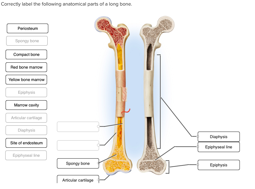
The endosteum (plural endostea) is a thin vascular membrane of connective tissue that lines the inner. Correctly label the following anatomical parts of a long bone. The endosteum is located on the internal surface of the bone, being the membranous layer that covers the medullary cavity, the bony trabeculae (spongy part of the bone), the haversian canals and internal walls of the compact long bones. Long bones are found in the arms (humerus, ulna, radius) and legs (femur, tibia, fibula), as well as in the fingers (metacarpals, phalanges) and toes (metatarsals, phalanges). The diaphysis and the epiphysis. The other encases the inside, and is called called the endosteum.these sheaths or membranes consist of a material called connective tissue. One encompasses the outside, and is called the periosteum; The diaphysis is the hollow, tubular shaft that runs between the proximal and distal ends of the bone. Terms in this set (12). Spongy bone makes up most of the structure of short, flat and irregular bones and the inside of the epiphysis of long bones. Red marrow fills the spaces in some bones. (b) in this micrograph of the osteon, you can clearly see the concentric lamellae and central canals. There are three different types of endosteum.
The name of each type refers to the area of the inner bone where it is located long bone diagram. The ends of a long bone contain spongy bone and an epiphyseal line.
Share :
Post a Comment
for "Long Bone Diagram Endosteum ~ General features of a LONG BONE - Biology 225 with Watson at McNeese State University - StudyBlue"


Post a Comment for "Long Bone Diagram Endosteum ~ General features of a LONG BONE - Biology 225 with Watson at McNeese State University - StudyBlue"